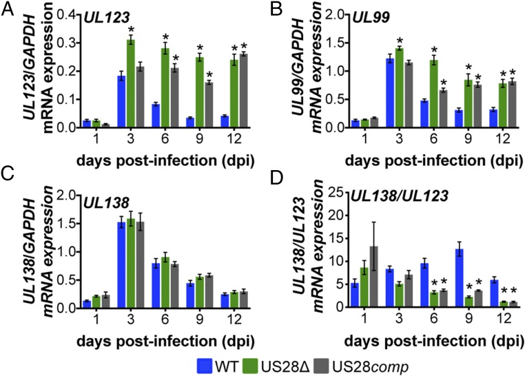
Fig. 5.
pUS28 represses UL123 and UL99 expression in infected Kasumi-3 cells at early times of latent infection. Kasumi-3 cells were infected with WT (blue), US28Δ (green), or US28comp (gray) (moi = 1.0) and sorted for mCherry-positive cells at 1 dpi. Cells were then harvested at the indicated dpi. (A) UL123, (B) UL99, (C) UL138, and (D) the ratio of UL138/UL123 expression were measured by RT-qPCR. Samples were normalized to GAPDH and analyzed in triplicate. Errors bars indicate SD, and statistical significance was calculated using two-way ANOVA and Dunnett’s post hoc analysis relative to WT virus at each time point; *P < 0.05.

