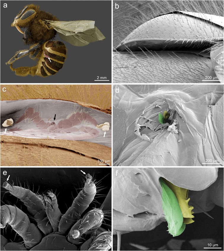
Fig. 2.
Feeding site of Varroa on adult honey bee imaged via low-temperature scanning electron microscopy. Images representative of 10 worker bees with attached mites prepared for imaging of which all 10 showed a wound in the intersegmental membrane. (A–F) Representative images of 10 bees parasitized by Varroa. Location of the mite shown with white arrow (A). The mite is wedged beneath the third tergite of the metasoma (B). When removed, a detailed impression of the mite can be observed in the intersegmental membrane in addition to a wound where the mouthparts of the mite would be (black arrow in C). Note, the ambulacra, or foot pads, of the mite (white arrows) remained attached to the membrane when the mite was extracted (C and D). Higher magnification of the wound reveals distinct grooves in the wound matching the modified chelicera of the mouthparts of the mite, colorized for clarity [moveable digit (yellow), corniculus (green)] (F). (A and C) Reproduced with permission from ref. 47.

