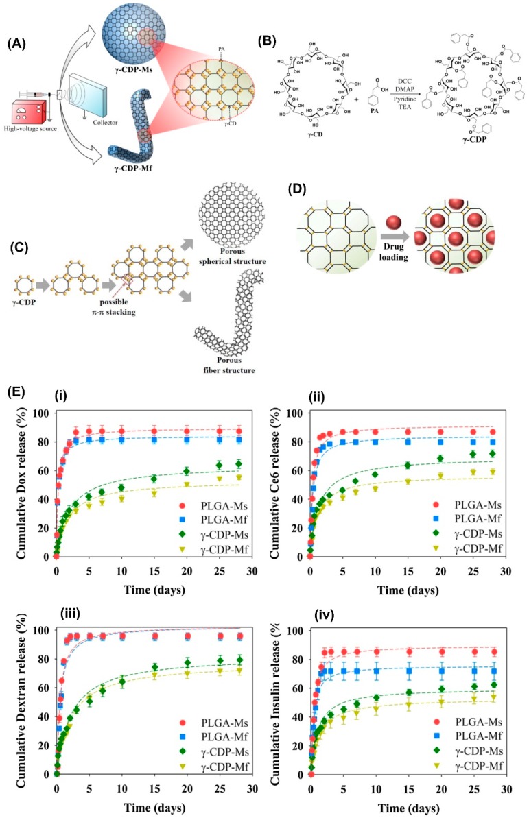
Figure 8.
(A) A schematic representation of the electrospinning and electrospraying of γ-CyDPs. (B) The synthesis pathway of γ-CyDP. (C) Cartoon schemes of γ-CyDP-microspheres (Ms) or γ-CyDP microfibers (Mf) with porous structure and (D) their drug loading. (E) The cumulative molecule (i) Dox, (ii) Ce6, (iii) dextran, and (iv) insulin release (wt. %) from PLGA-Ms, PLGA-Mf, γ-CyDP-Ms, and γ-CyDP-Mf (n = 3). The figure was reproduced from [182] with the permission of Elsevier, 2018.

