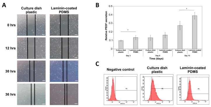
Figure 4.
RPE cells cultured on laminin-modified PDMS film demonstrate normal RPE biological functions. (A) Cell migration (wound healing) assay performed with hiPSC-RPE cells grown on plastic and laminin-modified PDMS. Scale bar = 100 µm (B) PEDF secretion by hiPSC-RPE cells grown on plastic and laminin-PDMS analyzed by ELISA assay in a time course of 3, 8 and 15 days. The values are the means from three independent measurements with SD error bars, * indicates statistically significant difference (ANOVA, p < 0.05). (C) Flow cytometry analysis of phagocytosis in populations of hiPSCs-RPE cells grown on plastic and laminin-PDMS.

