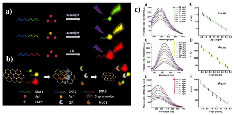
Figure 7.
(a) Schematic illustration of synthesizing DNA-Ag NCs and DNA-Ag/Au NCs; (b) schematically demonstrating the mechanism for assaying MUC1, CEA and CA125; (c) fluorescence spectra of the assaying model in the presence of varying concentrations of MUC1 (A), CEA (C) and CA125 (E), the linear relationships between F-F0 and the concentrations of MUC1 (B), CEA (D) and CA125 (F), respectively. Reproduced from [120], with permission from Elsevier, 2018.

