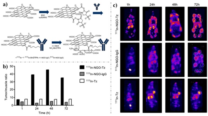
Figure 10.
(a) Synthesis of 111In-NGO-IgG or 111In-NGO-Tz. (b) Quantification of the biodistribution of the mice in c. (c) Representative whole-body SPECT images (MIPs) of the biodistribution of 111In-NGO-Tz, 111In-NGO-IgG, or 111In-Tz in spontaneous tumor-bearing BALB/neuT mice at 1, 24, 48, or 72 h i.p. injection. Tumors are located in mammary fat pads, with metastases in the axial and inguinal lymph nodes (white dashed circle in 72 h images). Tumor burden is likely different in each animal due to the nature of the spontaneous tumor model. Reproduced from [179], with permission from Elsevier, 2013.

