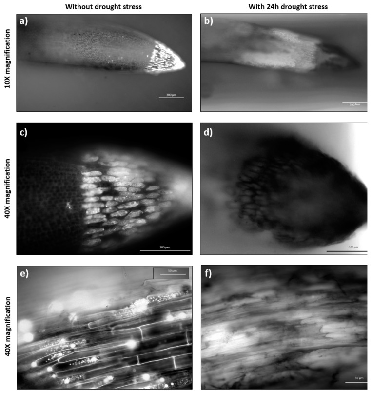
Figure 5.
Root tip and maturation zone under osmotic stress. Root in the microfluidic device after 72 h growth, under standard and 24 h stress conditions by 20% PEG; (a,b) show the root cap samples with 10× magnification; (c,d) show the root cap samples with 40× magnification; (e,f) shows the maturation zone with 40× magnification. The images were taken with an Axio Vert.A1 inverted microscope by Carl Zeiss (Germany).

