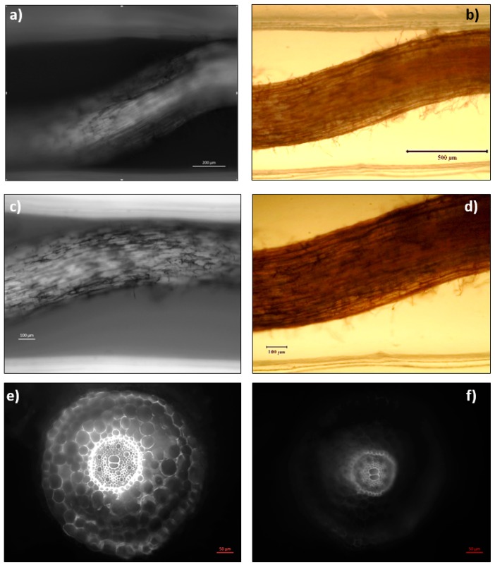
Figure 7.
Neutral red staining of root seen under light and fluorescent microscopy. Neutral Red staining of the root with (a,c) and without (b,d) fluorescence visualization was seen after 24 h, osmotic stress mostly concentrated in the internal vascular tissue. However, a reduction in the fluorescence was observed after 24 h stress in all samples. Cross-section images of elongation zone from a control sample (e) and the sample under 24 h osmotic stress (f). Elongation zone sample was sectioned 7 mm away from the root tip. The images “a, c, e and f” were taken with an Axio Vert.A1 inverted microscope by Carl Zeiss (Germany), and the images “b” and “d” were taken with the Nikon stereomicroscope (Japan).

