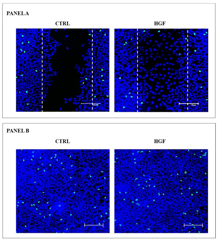
Figure 4.
pHH3+ immunofluorescence in wound-healing assay. Panel A: Maximum projection of representative optical spatial series with step size of 1 µm recovered in the area of the wound. The images show NT2D1 cells, cultured for 48 h with or without HGF. Phospho-Histone H3 positive cells were immuno-labelled in green (FITC signal), whereas cell nuclei were stained with TOPRO-3 and appear blue (scale bar: 150 µm). The dotted lines indicate the presumptive area of the wound (that is 263,486 µm2), calculated at the beginning of the wound-healing assay. Each whole photographic-field measures 562,500 µm2. All experiments were performed in triplicate. Panel B. Maximum projection of representative optical spatial series of the same samples of Panel A recovered in areas far from the wound in which cells appear over-confluent. Each photographic-field measures 562,500 µm2. Phospho-Histone H3 positive cells were immuno-labelled in green (FITC signal), whereas cell nuclei were stained with TOPRO-3 and appear blue (scale bar: 150 µm).

