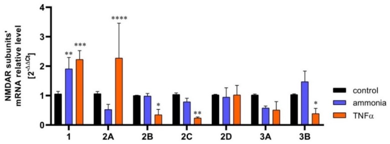
Figure 1.
The content of mRNA coding for NMDAR subunits in mouse cortical astrocytes cultured in control conditions and exposed for 72 h to 5 mM ammonia or 50 ng/ml TNFα treatment. Real-time PCR procedure was exactly as in Skowrońska et al., (2019) [72]. The mRNA expression was determined by TaqMan Gene Expression Assay (Applied Biosystems). The assay IDs were Mm00433790_m1 for GluN1 (Grin1); Mm00433802_m1 for GluN2A (Grin2a); Mm00433820_m1 for GluN2B (Grin2b); Mm00439180_m1 for GluN2C (Grin2c); Mm00433822_m1 for GluN2D (Grin2d); Mm01341723_m1 for GluN3A (Grin3a); Mm00504568_m1 for GluN3B (Grin3b); and Mm00607939_s1 for endogenous control β-actin. Results are mean ± SD (n = 3). (*) p < 0.05 vs control; (**) p < 0.01 vs control; (***) p < 0.001 vs control; (****) p < 0.0001 vs control; Two-Way ANOVA with Dunnett post-hoc test.

