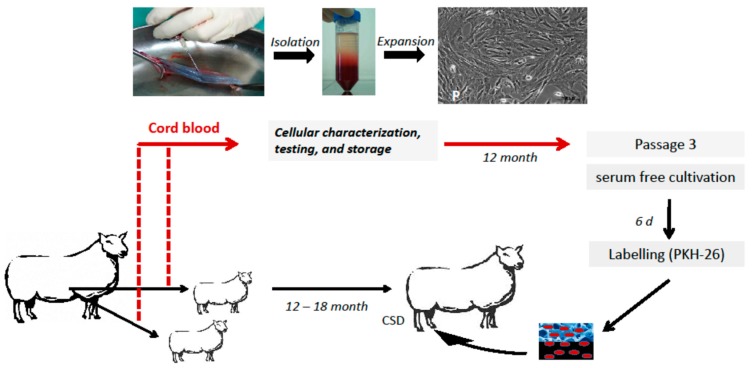
Figure 7.
Experiment design of the test group: autologous implantation of labeled USSC into the tibial bone defect. Ovine USSC were obtained, isolated; expanded, characterized by flow cytometry, and frozen. After 12 months, full-size 2.0 cm mid diaphyseal bone defects were created in the right tibia of adult sheep. Autologous USSC of the third passage were thawed, labeled with the membrane dye PKH-26 and 2 × 10⁷ cells were seeded onto four hydroxyapatite (HA) blocks and implanted into the defect.

