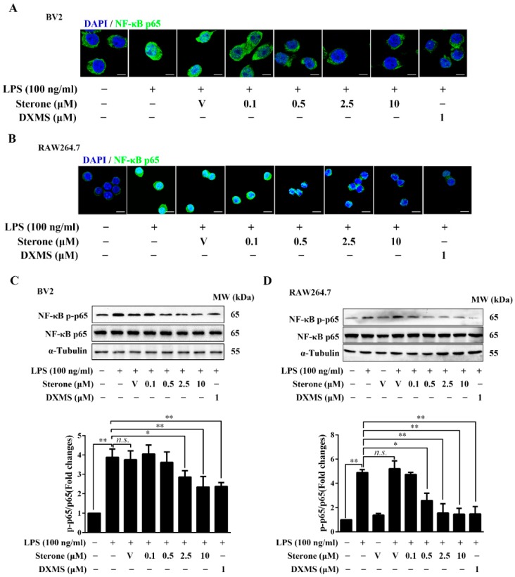
Figure 3.
Sterone inhibited LPS-induced NF-κB activation in BV2 cells and RAW264.7 cells. Cells were pre-treated with the indicated concentrations of sterone or DXMS for 30 min before the stimulation by 100 ng/mL LPS for 30 min individually. The nuclear localization of NF-κB p65 in BV2 cells (A) and RAW264.7 cells (B) were measured using immunofluorescence stain with an NF-κB p65 antibody (Green) and nuclear stain by 4′,6-Diamidino-2-phenylindole (DAPI, blue). The phosphorylation levels of NF-κB p65 in BV2 cells (C) and RAW264.7 cells (D) were measured using western blotting. V, Vehicle (HP-β-CD). Scale bars, 10 μm. Values are mean ± SD of three independent experiments. n.s, no significance. * p < 0.05, ** p < 0.01.

