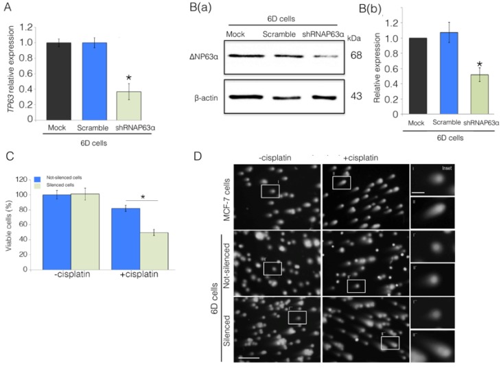
Figure 2.
ΔNp63α plays a role in the IL-1β induction of cisplatin resistance in 6D cells. Cells were transfected with empty vector (Mock), non-specific short hairpin RNA (Scramble), and the specific silencing RNA (shRNAp63α). (A) TP63 expression was evaluated by qPCR. Results represent the average of three independent experiments ± SD. Asterisks correspond to p = 0.001 relative to the controls, Mock and Scramble. (B(a)) Representative Western blot of ΔNp63α protein levels in the 6D cells. (B(b)) Densitometric values corresponding to ΔNp63α levels in (B(a)) were normalized to those of β-actin. (C) Cell viability levels determined in ΔNp63α-silenced and non-silenced cells in the absence or presence of cisplatin. Data represent the average of four independent experiments ± SD. Asterisks indicate significance relative to 6D cells at p = 0.001. (D) Comet assay to evaluate DNA integrity and damage by cisplatin in MCF-7, non-silenced, and shRNAp63α-silenced 6D cells. Cells not treated and treated with cisplatin were mixed with low-melting point agarose, lysed, and subjected to electrophoresis, followed by staining with ethidium bromide. DNA was visualized by fluorescence microscopy; scale bar = 100 μm. The insets in the right panel show magnified images taken from each condition; scale bar = 25 μm.

