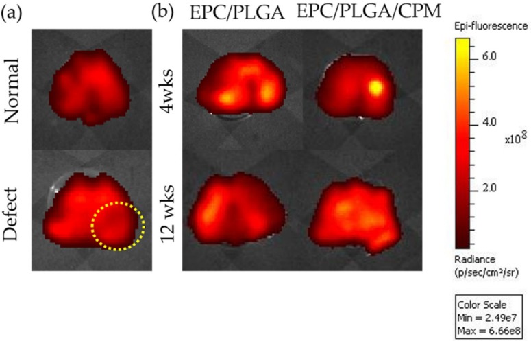
Figure 2.
Location of endothelial progenitor cells (EPCs) after transplantation for the normal and defect only groups; the yellow dotted circle indicates the defect site (a). Localization of EPCs by CM-Dil 4 and 12 weeks after PLGA scaffold transplantation with or without continuous passive motion (CPM) intervention (b). The bioluminescence was determined by in vivo imaging system (IVIS) measurements in the dissected tissue of the femurs of the EPC/PLGA and EPC/PLGA/CPM groups.

