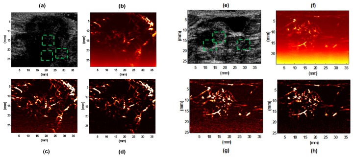
Figure 6.
B-mode image of invasive ductal carcinoma (IDC) type III breast lesion (a) and IDC type II breast lesion (e); their gross vasculature SVD images (b,f), respectively; their vasculature images after SVD+TH (c,g), respectively; final images of vasculature after applying the proposed denoising method (NLM+TH) (d,h), respectively.

