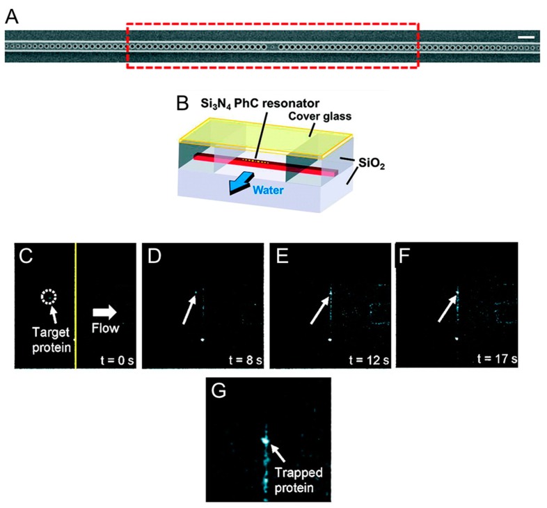
Figure 2.
Trapping of individual proteins with a photonic crystal resonator. (A) Scanning electron microscope image of the silicon nitrite photonic crystal resonator fabricated by Chen et al. [41] to trap nanometer-sized objects. The resonator consists of a central hole flanked by 53 holes on both sides and is operated at 1064 nm. Scale bar: 1 µm. (B) Schematic of the flow chamber used to flow protein molecules near the resonator. (C–F) Fluorescence microscope images of Cy5-labeled Wilson disease proteins inside the flow chamber. As the flow is activated (t = 0 s), the proteins that are conveyed near the optically excited resonator remain stably trapped. A high-resolution picture of a group of trapped proteins is shown in (G). Adapted with permission from [41]. Copyright 2012 American Chemical Society.

