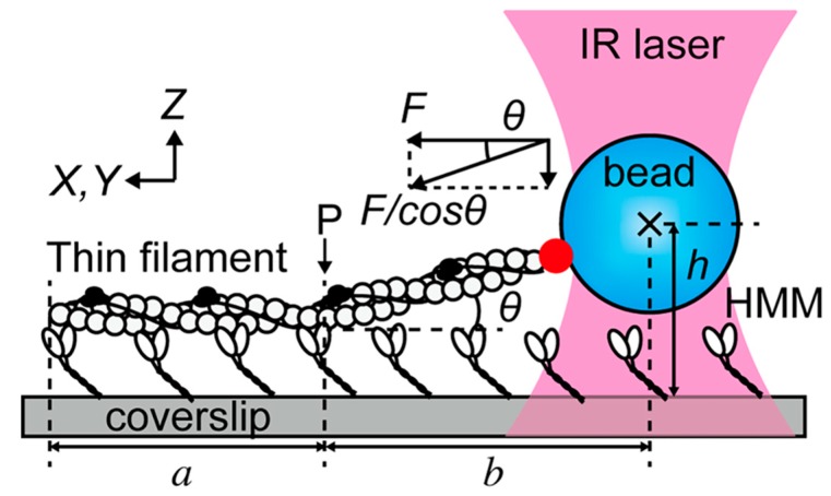
Figure 5.
Experimental setup used by Ishii et al. [91]. The filament made of actin, tropomyosin, and troponin is attached to the coverslip for a length a up to the point P by its interaction with heavy meromyosin molecules (HMM). One end is connected to a polystyrene bead trapped by an infrared (IR) laser. The filament is labeled with fluorophores and imaged using widefield techniques. Optical tweezers measure the intensity of the force F/cos θ, while fluorescence allows to estimate both the lengths h and b; in this way, the z-component of the force can be reconstructed. Reprinted with permission from [91].

