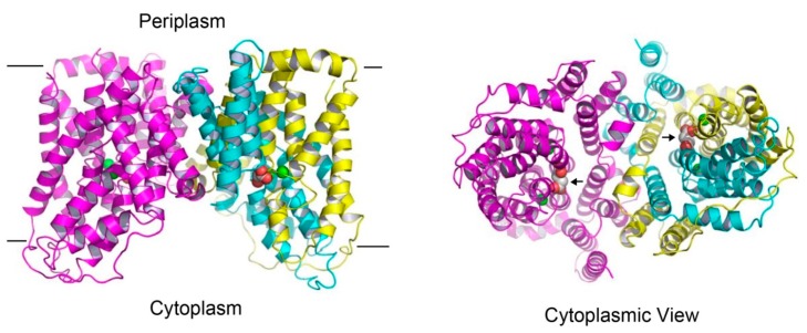
Figure 3.
Structure of the succinate-bound VcINDY. Structure of dimeric VcINDY, as viewed from the membrane bilayer (left panel). VcINDY is shown in ribbon rendition, the N (amino acids 18-231) and C (amino acids 232-462) domains in one protomer are colored cyan and yellow, respectively, whereas the other protomer is colored magenta. Na+ ions (green) and succinate are drawn as spheres. The cytoplasmic view of the VcINDY structure (right panel), highlighting the solvent-accessible succinate (black arrows) and buried Na+ ions. As such, the crystal structure captures the transporter in the inward-open state. Unless noted otherwise, structural analysis in this review was performed by using the program O and figure was prepared by using the software PyMOL.

