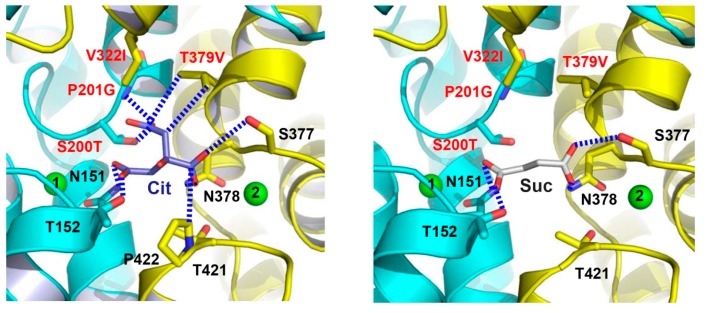
Figure 6.
The citrate- and succinate-binding sites in humanized variant of VcINDY. Structure of the citrate-binding site (left panel). Close-up view of the succinate-binding site (right panel). Citrate (light blue), succinate (grey) and relevant amino acids are drawn as stick models, while the Na+ ions are shown as green spheres. Humanizing amino-acid substitutions are highlighted in red. Dashed lines (blue) indicate the interactions between MT5 and citrate or succinate.

