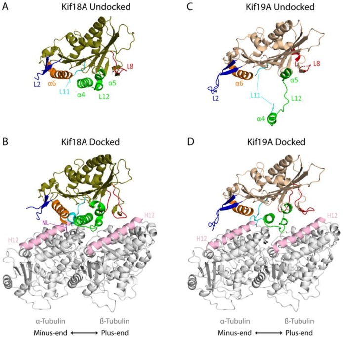
Figure 2.
Crystal structures of Kinesin-8 proteins and their cryo-EM MT docked state: (A) Undocked Kif18A complexed with Mg2+-ADP (PDB: 3LRE) [82]. (B) Nucleotide free Kif18A docked to the MT (PDB: 5OAM) [77]. (C) Undocked Kif19A complexed with Mg2+-ADP (PDB: 5GSZ) [79]. (D) Nucleotide free Kif19A docked to the MT (PDB: 5GSY; tubulin coordinates provided by N. Hirokawa) [79]. Key elements required for MT binding and/or MT destabilizing activity are color coded: Switch II cluster (α4-L12-α5) is shown in green, loop L2 in blue, helix α6 in orange, loop L11 in cyan, loop L8 in red, and neck linker (NL) in purple. Helix 12 of α- and β-tubulin is shown in light pink.

