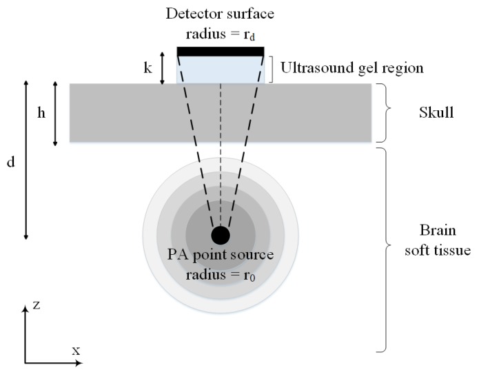
Figure 2.
2-D illustration of a simple model of skull used in our simulations. The model consists of a skull bone with a thickness of h located above the brain tissue. The spherical PA imaging target with the radius of is located at a depth of d below the skull layer and a flat ultrasound transducer with an element diameter of is remained in contact with the outer-skull surface and the coupling gel.

