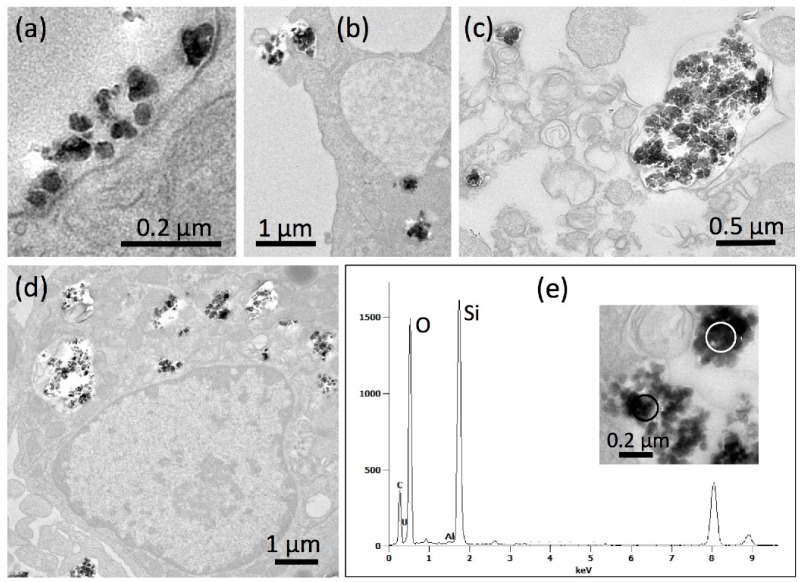
Figure 5.
Detection of precipitated P-6 silica nanoparticles in NR8383 cells by transmission electron microscopy (TEM). Cells were treated with 67.5 µg P-6 per mL in serum-free F-12K and fixed after 90 min (a,b) and 16 h (c–e). (a) P-6 NP attached to the cell; the underlying cell membrane appears undamaged. (b) Early stage of phagosome formation (upper left). (c) A P-6-filled phagolysosome released from a deteriorated cell. (d) NR8383 macrophage filled with numerous P-6-laden phagosomes. (e) Energy dispersive X-ray analysis (TEM-EDX) of a P-6-containing phagosome; white circle (inset) marks the analyzed area; results from black circle were highly similar (not shown). Prominent signals (in arbitrary units) were obtained for silicon (Si) and oxygen (O) at typical positions (in keV) of the spectrum.

