Figure 6.
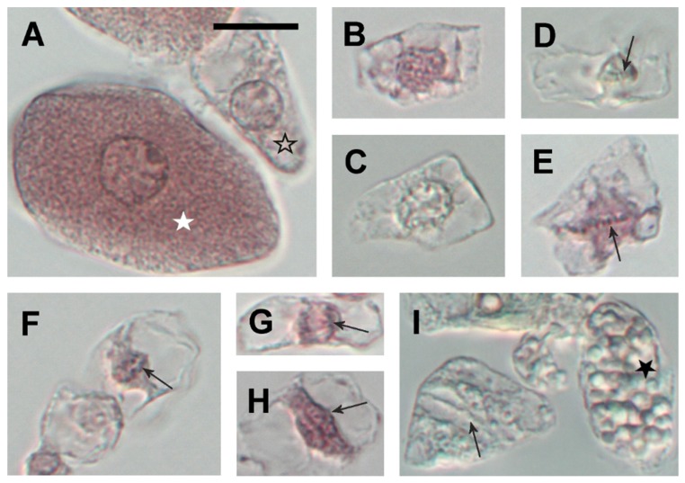
Cell types separated from L. japonicus root nodules after maceration in pectinase. Infected cell (white star) and noninfected interphase cell (open star) (A). Early and late prophase cells, respectively (B,C). Metaphase cells (arrows point to the metaphase plate) (D,E). Anaphase cell (arrow points to one group of chromatids) (F). Early and late telophase cell (arrows point to one of the twin nuclei) (G,H, respectively). Cell in cytokinesis (arrow points to the cell plate) and noninfected fully differentiated cell with large starch grains (black star) (I). Images (A,B,E–H): cells stained with cold acetocarmine, images (C,D,I): unstained. All images are in the same magnification, bar = 20 µm.

