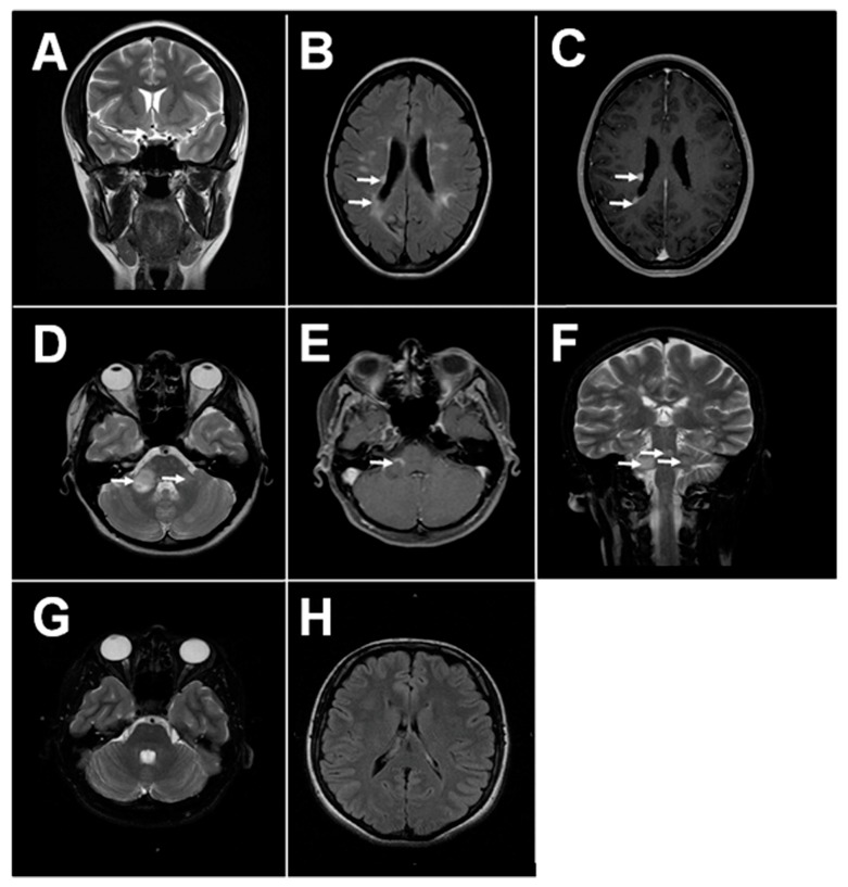
Figure 1.
Magnetic resonance imaging (MRI) of MS and health brain. (A) Typical MS lesions in the right optical nerve on coronal T2 image; (B,C) Typical MS lesions in periventricular white matter on axials T2 and T1 post contrast images, respectively; (D–F) Typical MS lesions in the white matter of sub-tentorial structures (pons, right middle cerebellar peduncle): on axials T2 and T1-post-contrast, and coronal T2 images, respectively; (G,H) MRI of the healthy adult brain: normal sub-tentorial structures on axial T2 images, normal periventricular white matter on axial FLAIR images, respectively.

