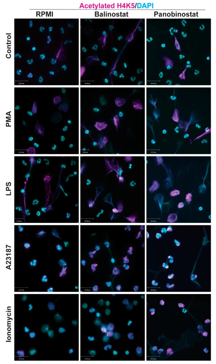
Figure 1.
Confocal microscopy images showing histone deacetylase inhibitors (HDACi) promote histone acetylation. Neutrophils were treated with negative control (RPMI media), or NETotic agonists (25 nM PMA; 4 μM A23187; 5 μg/mL lipopolysaccharides (LPS) from E. coli 0128; 5 μM Ionomycin) for 120 min. Cells were then fixed, immunostained, and imaged for histone acetylation (H4K5ac) and DNA (DAPI). Cells treated with RPMI show typical polymorphonuclear morphology of neutrophils. When treated with HDACis, belinostat and panobinostat, neutrophils show a further increase in histone acetylation. Blue, DAPI staining for DNA; Magenta, H4K5ac. Scale bar, 14 µm. n = 2–3. See Supplementary Figures S1–S3 for single channel confocal images.

