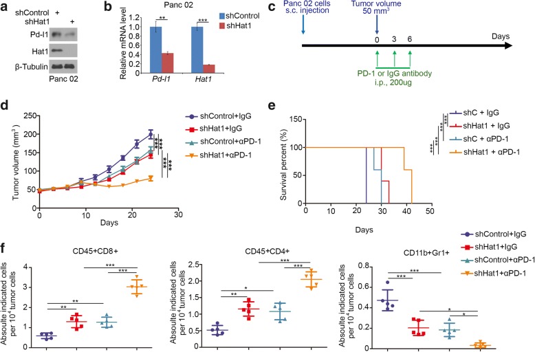
Fig. 5.
Knockdown of HAT1 improves the efficacy of immune checkpoint blockade by decreasing the PD-L1 expression in vivo. a and b, Panc 02 cells were infected with lentivirus vectors expressing control or Hat1-specific shRNAs. Forty-eight hours postinfection, the cells were harvested for Western blotting (a) and RT-qPCR analysis (b). The data shown are the mean values ± SD from three replicates. **, P < 0.01; ***, P < 0.001. c, Schematic diagram depicting the treatment plan for mice bearing subcutaneous Panc 02 tumors. d, Panc 02 cells were infected with lentivirus vectors expressing control or Hat1-specific shRNAs. Seventy-two hours after selection with puromycin, 5 × 10⁶ cells were injected subcutaneously into C57BL/6 mice. Mice (n = 5/group) were treated with anti-PD-1 (200 μg) or nonspecific IgG for 42 days. The growth curves of tumors with the different treatments are shown in (d). e, Kaplan-Meier survival curves for each treatment group demonstrate the improved efficacy of combining PD-1 mAb with HAT1 knockdown. ***P < 0.001. (Gehan-Breslow-Wilcoxo test). f, At the end of the treatment, the numbers of infiltrated CD45+CD8+ T cells, CD45+CD4+ T cells, and CD11b+Gr1+ myeloid cells that infiltrated tumors following the different treatments were analyzed by FACS. All data are shown as the mean values ± SD. ns, not significant, * P < 0.05, ** P < 0.01, *** P < 0.001

