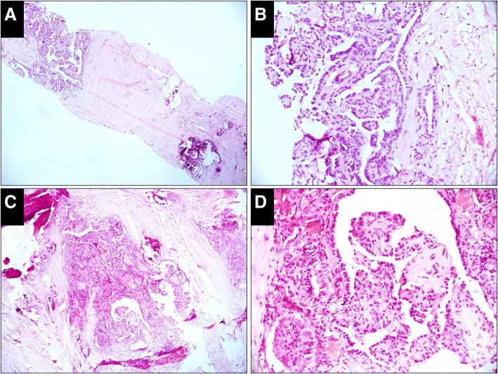
Fig. 6.
Case classified as malignant in both the CNB and resected specimens (H&E). a, Papillary pattern with calcification is embedded in the hyalinized collagen (× 50). b, High-power view of the papillae shows the cells with typical nuclear features of papillary thyroid carcinoma (× 200). c, An infiltrating tumor with stromal fibrosis is prominent in the resected specimen. The major architecture of the tumor is a papillary and elongated glandular pattern (× 50). d, High-power view of the papillae shows the cells with the nuclear features of papillary thyroid carcinoma (× 200)

