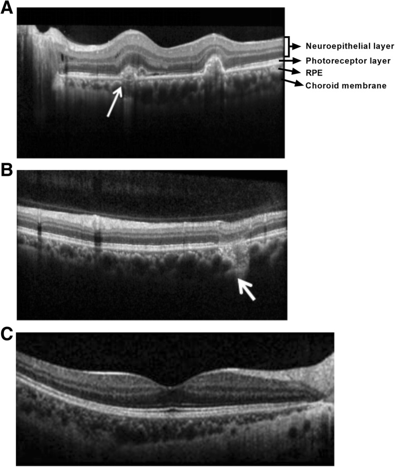
Fig. 5.

Typical OCT images of the MFC eyes. a The CNV secondary active inflammation lesion at the macula nasal elevates subretinally with a rough border and a few subretinal effusion (white arrow). b RPE-deficiency and choroidal scar formation of the inactive inflammatory lesion can be detected at the supraorbital vascular arch (white arrow). c The healthy control
