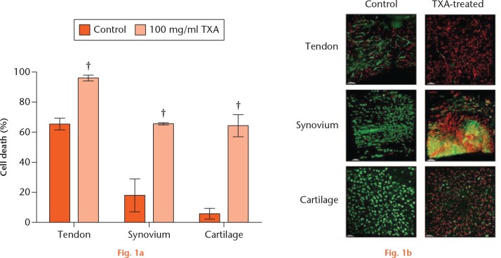
Tranexamic acid (TXA) toxicity in periarticular tissues ex vivo. a) Percentage of dead cells in control (0 mg/ml) and TXA-treated (100 mg/ml) cells in each periarticular tissue (n = 3 tendon, synovium; n = 4 cartilage) after 16 hours of treatment. Mean ± standard error of the mean; Student’s paired t-test. †p < 0.01. b) Tendon, synovium, ligament, and cartilage tissue imaged by confocal microscopy with 5-chloromethylfluorescein diacetate (CMFDA) and propidium iodide (PI) stain at 16 hours of treatment in control and TXA-treated (100 mg/ml TXA cells). Green stained cells counted as live and red stained cells counted as dead.

An official website of the United States government
Here's how you know
Official websites use .gov
A
.gov website belongs to an official
government organization in the United States.
Secure .gov websites use HTTPS
A lock (
) or https:// means you've safely
connected to the .gov website. Share sensitive
information only on official, secure websites.
