
Figure 4.
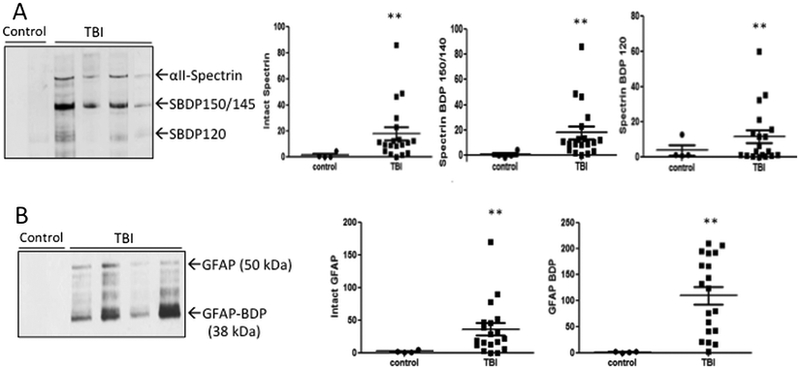
Presence of neuronal and glial proteolytic biomarkers αII-Spectrin and its breakdown products (SBDPs) and GFAP and its BDP in MV/E samples isolated from control and TBI CSF. (A) Immunoblots images showing the presence of these markers in TBI CSF isolated MV/E. (B) quantification of levels of these protein markers in MV/E preparations isolated from TBI vs. control CSF. **p < 0.01 (statistical significant). For standardization, each lane was loaded with protein from 3 ×10⁸ MV/E particles. Sample size: control n=4, TB, n= 19.