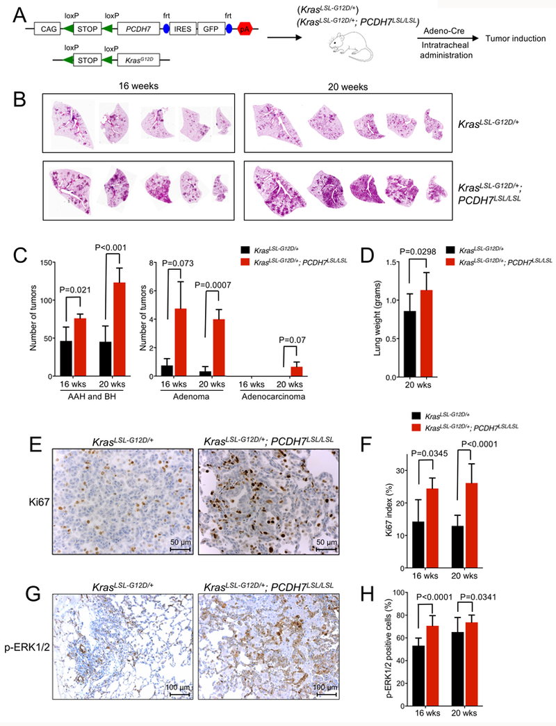
Figure 1. PCDH7 accelerates lung tumorigenesis in the KrasLSL-G12D model.

A, Schematic depicting the PCDH7LSL and Kras LSL-G12D transgenes and overview of experimental design. To induce lung tumors, KrasLSL-G12D; PCDH7LSL/LSL or control KrasLSL-G12D mice were infected with Adeno-Cre through intratracheal administration. B, H&E staining of lung lobes harvested at 16 and 20 weeks post-infection. Five independent lobes from one animal of each genotype are shown. C, Tumor burden analysis at 16 and 20 weeks post-infection. n=5–6 mice/group, 5 sections/mouse analyzed. Tumors were counted and classified into atypical adenomatous hyperplasia (AAH)/bronchiolar hyperplasia (BH), adenomas, or adenocarcinomas. D, Weight of lungs collected from KrasLSL-G12D; PCDH7LSL/LSL or KrasLSL-G12D mice at 20 weeks post-infection. E, IHC staining of Ki67 at 20 weeks and F, quantification of Ki67 index for lung sections harvested from KrasLSL-G12D; PCDH7LSL/LSL or control KrasLSL-G12D mice at 16 and 20 weeks post-infection. n=2–4 mice/group, up to 10 fields quantified/animal. G, IHC staining of pERK1/2 for lung sections from KrasLSL-G12D; PCDH7LSL/LSL or control KrasLSL-G12D mice at 16 weeks post-infection. H, quantification of p-ERK1/2 IHC staining at 16 and 20 weeks post-infection. n=2 mice/group, 7 fields quantified/animal.
