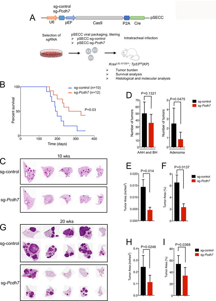
Figure 2. In vivo inactivation of Pcdh7 reduces lung tumor burden and prolongs survival of KrasLSL-G12D; Tp53fl/fl mice.

A, Schematic of in vivo CRISPR/Cas9 editing. KP mice were infected with a sg-control or sg-Pcdh7 through intratracheal administration of pSECC lentivirus expressing Cas9 and Cre. B, Survival analysis of KP mice infected with sg-control or sg-Pcdh7 lentivirus. C, H&E staining of lung lobes harvested at 10 weeks post-infection. D, Tumor burden analysis at 10 weeks post-infection. Tumors were counted and classified into AAH, BH, or adenomas based on histopathologic analysis. n=5–7 animals/group, 5 lobes/animal, 1 section/lobe analyzed. E, Total tumor area (inches2) at 10 weeks post-infection based on histopathologic analysis of lung sections using the NIH Image J program. F, Percent tumor area compared to total area of lung lobes at 10 weeks post-infection (determined using NIH Image J). G, H&E staining of lung lobes harvested at 20 weeks post-infection. H, Total tumor area (inches2) at 20 weeks post-infection based on histopathologic analysis of lung sections. I, Percent tumor area compared to total area of lung lobes at 20 weeks post-infection.
