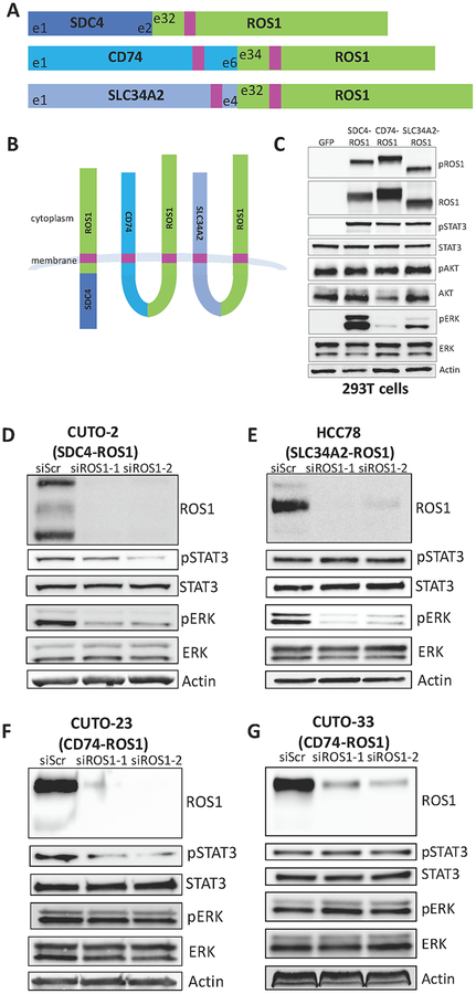
Figure 1. ROS1 fusion partners dictate differential activation of downstream signaling pathways.
(A) Diagram of the commonly occurring ROS1 fusion oncoproteins, which were studied here. Pink denotes a transmembrane domain. (B) Topological configuration of ROS1 fusions based on CCTOP computational analysis (28). (C) Immunoblot analysis of 293T cells transiently transfected for 48h with GFP, SDC4-ROS1, CD74-ROS1, or SLC34A2-ROS1, with 5h serum starvation. The pROS1 antibody used recognizes Y2274 of the full-length ROS1 protein. (D-G) Immunoblot analysis of patient-derived cell lines expressing (D) SDC4-ROS1, (E) SLC34A2-ROS1, or (F-G) CD74-ROS1 with siRNA-mediated knockdown of ROS1 (55h after transfection). Data shown in (C-G) are representative of ≥ 3 independent experiments.

