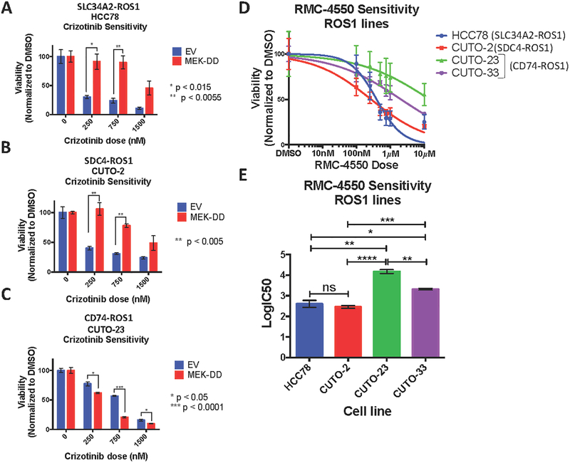
Figure 2. MAPK pathway signaling is necessary and sufficient for survival of SDC4-ROS1-positive and SLC34A2-ROS1-positive lines, but not a CD74-ROS1 positive line.
(A-C) Crystal violet quantification of ROS1 fusion-positive patient-derived cell lines (A) HCC78, (B) CUTO-2 and (C) CUTO-23, expressing empty vector or constitutively active MEK-DD, treated with DMSO or a dose-response of the ROS1 inhibitor crizotinib for 6 days. (D) Crystal violet quantification of HCC78 (SLC34A2-ROS1), CUTO-2 (SDC4-ROS1), CUTO-23 (CD74-ROS1), and CUTO-33 (CD74-ROS1) cell lines treated with DMSO or a dose-response of the SHP2 inhibitor RMC-4550 for 6 days. (E) Half-maximal inhibitory concentration (IC50) determination for the SHP2 inhibitor RMC-4550 in the indicated ROS1 patient-derived cell lines based on crystal violet quantification of the experiment in (D). Data represent three independent experiments. Data represented as mean +/− s.e.m.

