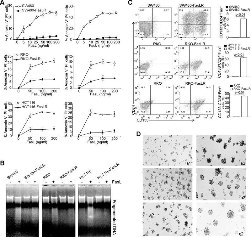
Figure 4. FasL selection enriches colon CSC-like cells.
A. SW480, RKO, HCT116, and the respective FasL-resistant cell lines as indicated were cultured in the presence of FasL at the indicated concentrations for 24h. Cells were stained with PI and annexin V. Early apoptosis (Annexin V+PI-) and apoptotic cell death (Annexin V+PI+) were quantified. B. The three pairs of parent and FasL-resistant cell lines were either untreated or treated with FasL (200 ng/ml) for 24h. Genomic DNA was isolated from the cells and analyzed by 1.5% agarose gel electrophoresis. C. The three pairs of parent and FasL-resistant cell lines were stained with CD133-, CD24-, and Fas specific mAbs and analyzed by flow cytometry. Shown are representative plots of CD133 and CD24 phenotypes (left two panels). CD133+CD24loFaslo cell subsets were then quantified and presented at the right panel. D. The parent and FasL-resistant cell lines were cultured in ultra-low attachment tissue culture plates with serum-free DMEM medium supplemented with EGF (20 ng/ml) and basic FGF (10 ng/ml), respectively, for 10 days. Shown are representative images of cell morphology of three independent experiments. a1 SW480, a2: SW480-FasLR, b1: RKO, b2: RKO-FasLR, c1: HCT116, C2: HCT116-FasLR.

