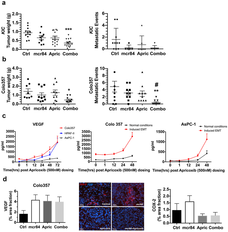
Figure 1. Combination therapy with apricoxib and mcr84 reduced tumor growth and metastasis in murine models of pancreatic cancer.
(a) At 3 weeks of age, KrasLSL-G12D; Cdkn2afl/fl; Ptf1aCre/+ (KIC) mice were randomized to receive saline (n = 11), mcr84 (n = 10), apricoxib (n = 13), or mcr84 plus apricoxib (n = 13). All mice were sacrificed when they were 7 weeks old. Mean tumor weight and metastasis burden were compared. (b) A total of 1106 Colo357 cells were injected orthotopically into NOD/SCID mice. Treatment began when established tumors were visible by ultrasound and consisted of control (n = 8), mcr84 (n = 10), apricoxib (n = 10) or mcr84 plus apricoxib (n = 10) and continued for 4 weeks, after which mean tumor weight and metastasis burden were shown. Data are displayed in a scatter plot with mean ± SEM. *P < 0.05, **P < 0.01, ***P < 0.005 vs. control; #P < 0.05 vs. single-agent mcr84 or apricoxib by ANOVA with Dunn’s MCT. (c) Human pancreatic cancer cell lines, HPAF-II, Colo357, and AsPC-1 were treated with 500 nM apricoxib and evaluated by ELISA for the production of VEGF. Colo357 and AsPC-1 were plated under normal conditions or conditions of forced EMT (50 ng/ml TGFβ on collagen I -coated plates for 24 hours). VEGF levels were evaluated by ELISA after 500 nM apricoxib treatment. Biological repeats have been performed (n=3) and data are displayed as mean ± SEM. Paraffin-embedded tumor sections from Colo357 tumor-bearing mice were analyzed for (d) VEGF and COX-2 expression by immunofluorescence. Quantification of percentage area fraction is shown. Data are displayed as mean ± SEM and represent 5 images per tumor with 3 animals per group. Representative images (COX-2, red; DAPI, blue) are shown for Colo357 tumors. Total magnification is 400X. Scale bars are presented as indicated.

