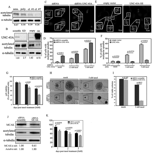
Figure 6. UNC-45A depletion exacerbates paclitaxel-mediated stabilizing effects on mitotic spindles and restores sensitivity to paclitaxel.
A. Western blot analysis for levels of UNC-45A acetylated α-tubulin in paclitaxel-sensitive and paclitaxel-resistant (polyclonal, clone #1, and clone #7) COV362 ovarian cancer cells. Numbers indicate the ratio between UNC-45A and α-tubulin. B. Left, Western blot analysis for levels of UNC-45A acetylated α-tubulin in COV362 ovarian cancer cells transduced with either shRNA scramble or shRNA-UNC-45A. Numbers indicate the ratio between acetylated α-tubulin and α-tubulin. Right, Western blot analysis for levels of UNC-45A in COV362 ovarian cancer cells infected with either empty vector (empty) or UNC-45A (overexpressing, OE). Numbers indicate the ratio between acetylated α-tubulin and α-tubulin. C. Mitotic figures containing multipolar spindles in either shRNA scramble or shRNA UNC-45A knockdown COV362 ovarian cancer cells in presence of 5 nM of paclitaxel as evaluated by γ-tubulin and DAPI staining. Arrows indicate spindle poles. Asterisks indicate cells with multipolar spindles. D. Quantification of cells containing multipolar spindles per each condition (mock: (−) n=20, (+) n=34; 1.5 nM paclitaxel: (−) n=23, (+) n=26; 5 nM paclitaxel: (−) n=29, (+) n=26). E. Mitotic figures containing multipolar spindles in either empty vector or UNC-45A overexpressing (OE) COV362 ovarian cancer cells in presence of 5 nM of paclitaxel as evaluated by α-tubulin and DAPI staining. Arrows indicate spindle pole. Asterisks indicate cells with multipolar spindles. F. Quantification of cells containing multipolar spindles per each condition (mock: (−) n=26, (+) n=37; 3 nM paclitaxel: (−) n=29, (+) n=31; 5 nM paclitaxel: (−) n=28, (+) n=32). G. Residual cell viability of shRNA scramble and shRNA-UNC45A knockdown COV362 cells exposed to 5nM paclitaxel over a period of 6 days. H. Equal numbers of shRNA scramble and shRNA-UNC- 45A SKOV-3 cells were seeded in soft agar for a period of 10 days prior paclitaxel treatment (5nM) over a period of three weeks. Per each condition, colonies were visualized using an inverted scope. Residual cell viability per each condition was evaluated in colonies’ biopsies via trypan-blue exclusion assay. All experiments were conducted in triplicates. I. Quantification of residual cell viability per each condition (mock: shRNA scramble n=83, shRNA UNC-45A n=75; 5 nM paclitaxel: shRNA scramble n=60, shRNA UNC-45A n=52). L. Western blot analysis for levels of MCAK and acetylated α-tubulin in siRNA scramble versus siRNA MCAK treated COV362 cells. Numbers indicate the ratio between MCAK and alpha-tubulin or acetylated alpha-tubulin and alpha-tubulin. M. Residual cell viability of siRNA scramble and siRNA-MCAK COV362 cells exposed to 5nM paclitaxel over a period of three days.

