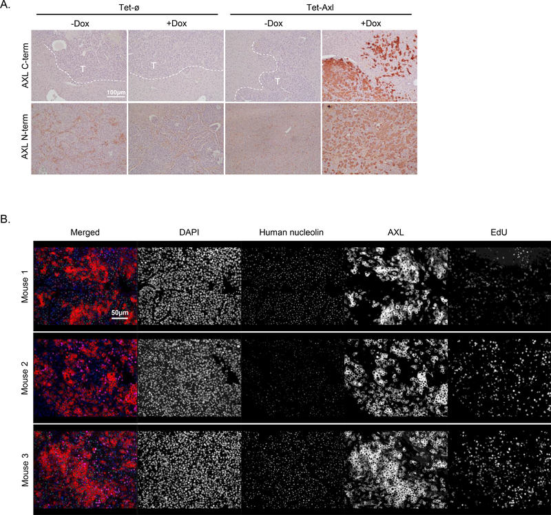
Figure 5.
AXL-on tumors are heterogeneous for AXL expression and EdU incorporation. A, Representative IHC staining to detect either the C-terminus or N-terminus of AXL in liver tumors from each group. Dashed white lines depict the border between tumor and liver tissue; ‘T’ indicates the tumor region (n = 3 per group; scale bar, 100 μm). B, Multiplex IF staining for DAPI, human nucleolin, AXL (C-terminus), and EdU incorporation in liver tumors from 3 different AXL-on mice; scale bar, 50 μm.

