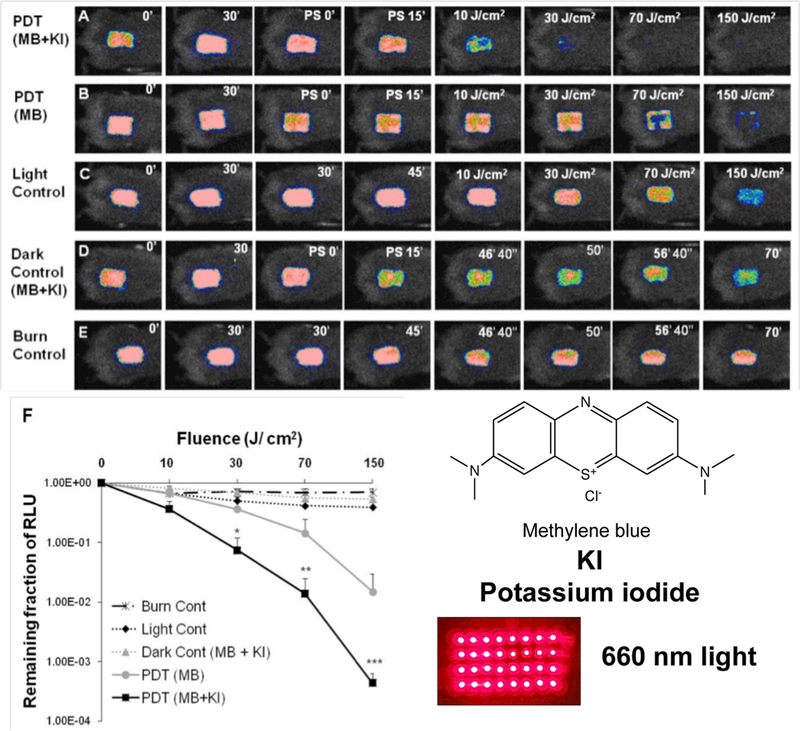
Figure 3. PDT mediated by MB plus KI in vivo.

(A-E) Successive bacterial bioluminescence images of representative mouse burns infected with 10(8) CFU of luminescent MRSA (USA 300) treated with: (A) PDT using mixture of MB (50 μM) + KI (10 mM) or (B) PDT using MB (50 μM) at 30 min after bacterial inoculation + 15 min from PS application. PDT was carried out with a combination of 50 μL of a mixture containing MB + KI or MB alone and 150 J/cm2 red light (660 ± 15 nm, 100 mW/cm2). (C) Light alone; (D) applied with mixture of MB + KI, but without red light illumination (dark control); (E) burn control without any treatment. (F). Dose-response plot of mean bacterial bioluminescence of mouse burns infected with MRSA (USA 300) after treatment with: light alone, mixture of MB (50 μM) + KI (10 mM) (dark control), PDT using MB (50 μM) alone or mixture of MB (50 μM) + KI (10 mM). From (45) no permission necessary.
