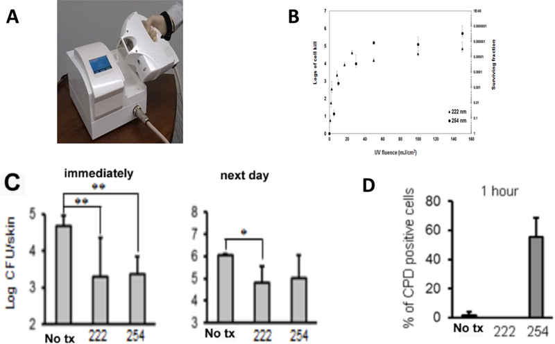
Figure 7. Comparison of 222 nm and 254 nm UVC.

(A) Photograph of 222 nm Kr/Cl excimer lamp. (B) Comparison of 222 nm and 254 nm UVC for in vitro inactivation of S. aureus on an agar plate. (C) S. aureus CFU extracted from a mouse wound either immediately or 24 hours after treatment with 750 mJ/cm2 of either 222 nm or 254 nm UVC. (D) % of CPD-positive cells quantified by counting 10 random high-power (×400) fields of tissue sections removed 1 hour post exposure to 150 mJ/cm2 of either 222 nm or 254 nm UVC. From (119) no permission necessary.
