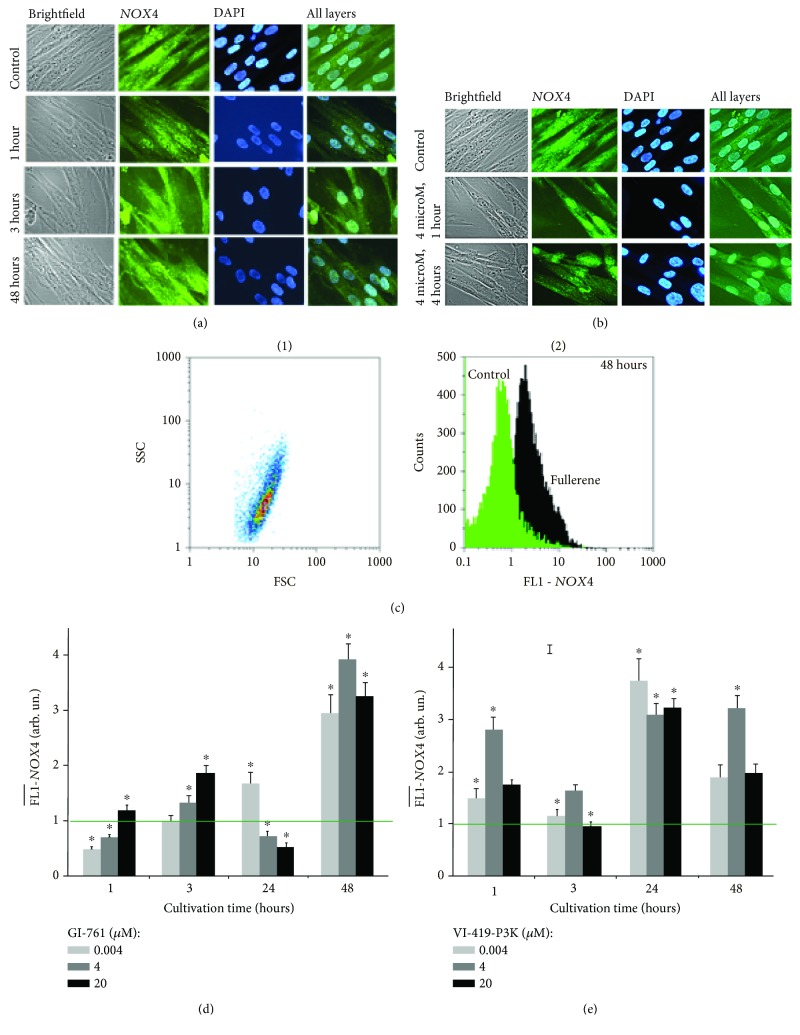
Figure 5.
NOX4 expression in HELFs incubated with GI-761 and VI-419-P3K. (a, b) Fluorescent microscopy: localization of NOX4 within fixed cells stained with DAPI and antibodies to NOX4 in HELFs treated with (a) GI-761 and (b) VI-419-P3K. (d, e) FCA: FL1-NOX4 levels for HELFs treated with GI-761 (d) and VI-419-P3K (e). Concentrations and times of exposure shown in graphs.

