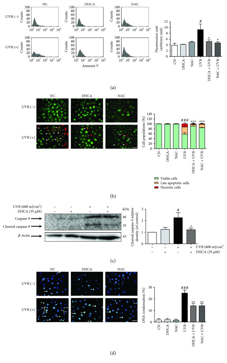
Figure 6.
Capacity of DHCA to decrease apoptosis features induced by UVB. L929 fibroblasts were pretreated with 35 μM DHCA or 1 mM NAC for 1 h, irradiated with UVB (600 mJ/cm2), and after 24 h, analyses were performed. (a) Cells were stained with annexin V, and apoptotic cells were quantified by flow cytometry. (b) Cells were double stained with acridine orange and propidium iodide, and viable cells (green fluorescence nuclei), late apoptotic cells (orange fluorescence nuclei, white headless arrow), and necrotic cells (red fluorescence nuclei, white arrows) were observed on fluorescence microscopy and quantitated. (c) Active caspase 9 expression was determined and quantified by western blot analysis using specific antibodies. (d) Cells were stained with Hoechst 33342 stain, and cells with condensed nuclei (white arrows) were observed on fluorescence microscopy and quantitated. Each column represents the mean ± SD (n = 3). ###p < 0.001 and #p < 0.05: significantly different from nonirradiated and nontreated cells (NC); ∗∗∗p < 0.001, ∗∗p < 0.01, and ∗p < 0.05: significantly different from irradiated and nontreated cells (UVB). Scale bars: 50 μm.

