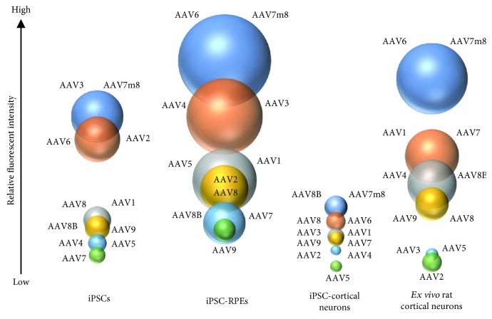
Figure 4.
Overview of AAV-eGFP transgene expression across four cell types. The illustration shows cell type and serotype comparison between induced pluripotent stem cells (iPSC), iPSC-derived retinal pigmented epithelium (RPE), iPSC-derived cortical neurons, and ex vivo-isolated rat cortical neurons. The size of the bubble is proportional to the level of GFP expression measure within and across cell type.

