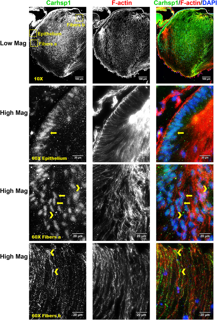
Fig. 6. Immunofluorescence analysis of expression of Carhsp1 in mouse lens.

Immunostaining for Carhsp1 (green), F-actin (red) and DAPI (nuclei, blue) in P0.5 CD1 mouse lens sections. Low magnification images show Carhsp1 is expressed in both lens epithelium and fiber. High magnification images demonstrate Carhsp1 is mostly found in the nuclei at lens epithelial as well as periphery fiber cells (see yellow arrows) while in the cytoplasm (see yellow chevrons) at anterior (apical) portions of fiber cells. Yellow boxes were used to indicate the positions of high magnification images, including epithelium, fibers position a (periphery fiber cells), and fibers position b (anterior/apical fiber cells). Scale bars: 100 µm (low magnification, top panels) and 20 µm (high mag, lower panels).
