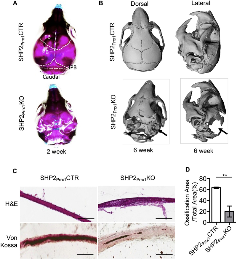
Figure 1. SHP2 ablation in the Prrx1+ mesenchymal cells impairs intramembranous ossification.

A. Representative skull images of 2-week-old SHP2Prrx1CTR and SHP2Prrx1KO mice stained with Alcian blue and Alizarin Red. n=3. B. μ-CT radiographs demonstrating the skull structure of 6-week-old SHP2Prrx1CTR and SHP2Prrx1KO mice. Note that parietal and interpariental bone ossification was defective (arrows) in the SHP2Prrx1KO mice, compared with age matched SHP2Prrx1CTR mice. n=3. C. Representative H&E (top) and von Kossa (bottom, counterstained by fast red) staining of murine parietal bone coronal frozen sections showing the appearance of a loose mesenchyme tissue and impaired mineralization of the parietal bone in the P0.5 SHP2Prrx1KO mice, compared with SHP2Prrx1CTR mice. Scale bar:100μm. D. Bar graphs show the quantitative data of parietal bone matrix mineralization in SHP2Prrx1CTR and SHP2Prrx1KO mice using NIH ImageJ software (n=3, **P<0.01, Student’s t-test).
