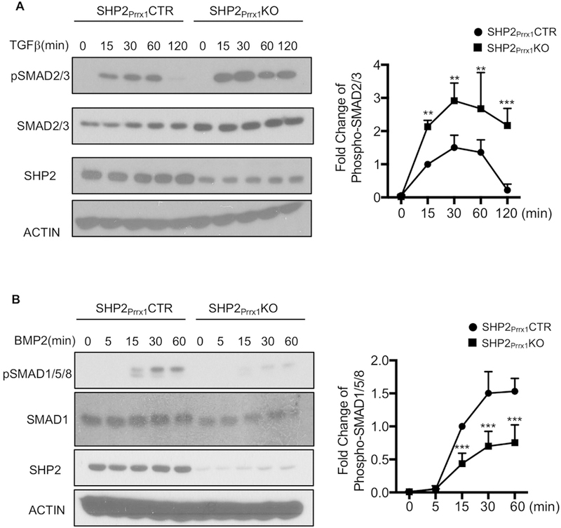
Figure 5. SHP2 differentially regulates TGFβ and BMP signaling pathways.

A. Immunoblotting data show that TGFβ-evoked SMAD2/3 phosphorylation was enhanced in the osteoblastic cells from SHP2Prrx1KO mice, compared to that from SHP2Prrx1CTR mice. Cells were starved overnight and stimulated with TGFβ (100ng/ml) for indicated time points. Phosphorylation of SMAD2/3 was quantified using NIH ImageJ software and normalized to the β-ACTIN controls, phosphorylation level of SMAD2/3 was corrected by the total level of SMAD2/3. (n=3, **p<0.01, ***p<0.001, Student’s t-test) B. Immunoblotting data showing decreased SMAD1/5/8 phosphorylation in response to BMP2 in SHP2Prrx1KO osteoblastic cells, compared to SHP2Prrx1CTR osteoblastic cells. Cells were starved overnight and stimulated with BMP2 (100ng/ml) for indicated time points. Phosphorylation of SMAD1/5/8 was quantified as described in A (n=3, **p<0.01, ***p<0.001, Student’s t-test).
