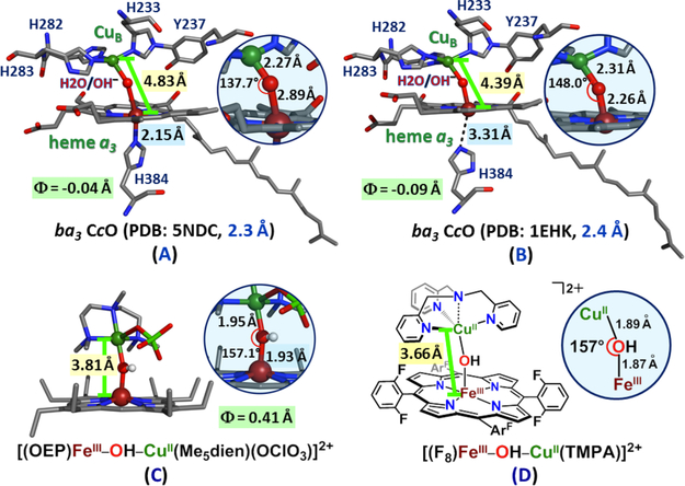
Figure 165.
X-ray structures of proposed μ-hydroxo (or μ-aquo) heme-copper centers proposed in CcO X-ray structures (A and B) or found in a small molecule model complex X-ray structure (C). For a different synthetic complex, [(F8)FeIII-(OH)-CuII(TMPA)]2+ with a tetradentate Cu center (D), structural parameters were determined from extended X-ray absorption fine structure (EXAFS) spectroscopy. Φ is the distance (above + , below −3) of the iron from plane of the porphyrin. Notably, the protein structure 5NDC (A) is said to be free of any possible damage derived from X-ray radiation, as it was determined utilizing an X-ray free electron laser (XFEL). Created using data from refs 987, 989, 1023, and 1071.

