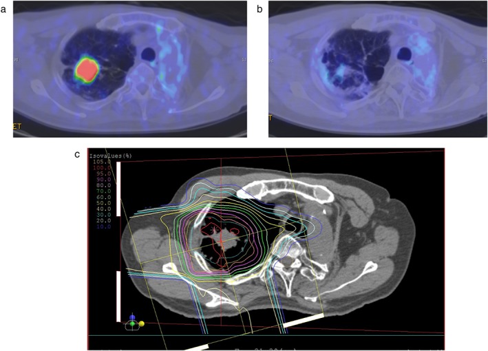
Figure 1.

The results of proton beam therapy after pneumonectomy in a 67‐year‐old squamous cell carcinoma patient. Positron emission tomography (a) before proton beam therapy and (b) nine months after treatment. (c) The dose distribution map of proton beam therapy for a lung cancer patient with idiopathic fibrosis. The region inside the innermost orange line received a 95% radiation dose, the pink line received a 90% radiation dose, and outer line is indicated by a 10% step.
