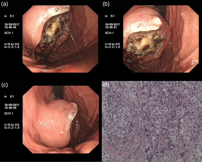
Figure 3.

(a,b) Electron gastroscopy: An oval tumor (3 cm × 4 cm) is shown in the fornix gastricus. (c) The surface of the tumor was unclear, covering the fossil moss and tissue brittle. (d) The pathological result: (fornix gastricus) solid cancer nest infiltration can be found in all six pieces of gastric mucosa and fibrous tissue.
