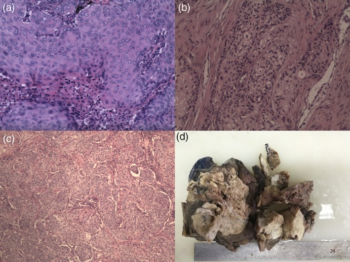
Figure 4.

(a) Intraoperative rapid frozen section (lung tumor): moderately differentiated squamous cell carcinoma (diameter 1.4 cm). (b) Intraoperative rapid frozen section (gastric tumor): non‐small cell carcinoma infiltration can be seen in the mucous membranes of the columnar epithelium (2.5 cm × 2 cm × 1 cm). (c) The postoperative pathology report: (i) lung tumor (7 cm × 6 cm × 7 cm), moderately differentiated squamous cell lung cancer; (ii) gastric tumor (diameter 6 cm), moderately differentiated squamous cell carcinoma; (iii) the results of immunohistochemistry: (lung tumor) CK7‐, TTF‐ 1‐, Napsin A‐, CK5 / 6 +, P63 +, P40 +, CD56‐, Syn‐, CgA‐, CK + and Ki‐67 indexes of approximately 30% +; (gastric tumor) CK7‐, TTF‐ 1‐, Napsin A‐, CK5 / 6 +, P63 + and Ki‐67 indexes of approximately 30% +. (d) Left lower lobe specimens (17 cm × 12 cm × 10 cm).
